

Loading article...
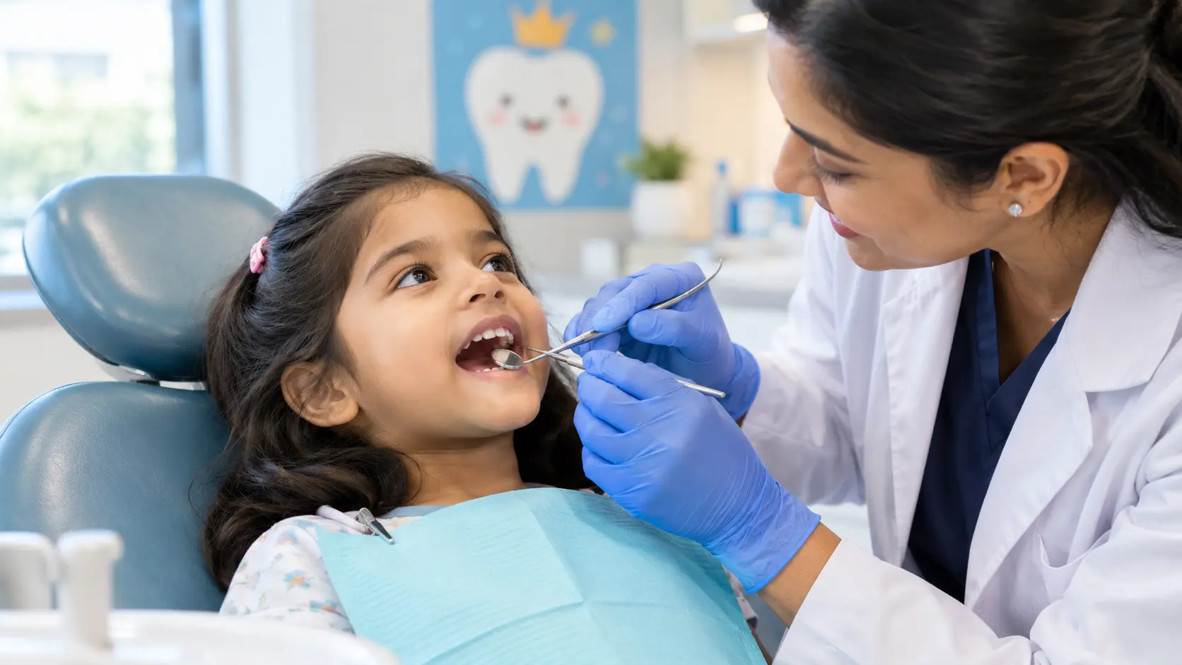
Tooth decay in children is one of the most common chronic conditions of early childhood, more common than asthma or allergies in many populations, yet it is also one of the most preventable. It begins quietly, often as a faint white spot on a tooth surface that a parent may never notice, and it can progress to pain, infection, and even tooth loss if left unaddressed. Globally, an estimated 514 million children have dental caries in their primary teeth, with the majority of that untreated decay concentrated in low and middle income countries. This guide explains what causes tooth decay in children, how to recognize it early, proven ways to prevent it, and the treatment options available when a cavity has already formed.
Every section below is written to give a direct, medically accurate answer first, so parents searching for urgent information, and AI-assisted search tools summarizing this topic, can find what they need quickly.
Tooth decay, also called dental caries, is the gradual destruction of tooth enamel and the deeper layers of the tooth caused by acid-producing bacteria in the mouth. When sugars and starches from food are left on the teeth, oral bacteria metabolize them and release acid, which slowly dissolves the mineral structure of the enamel. Over repeated cycles of acid exposure, this demineralization creates a cavity, a permanent hole in the tooth that will not heal on its own and needs dental treatment.
When this pattern of decay affects a child's primary teeth before the age of six, it is classified clinically as early childhood caries (ECC). According to the American Academy of Pediatric Dentistry, ECC is defined as the presence of one or more decayed, missing, or filled tooth surfaces in any primary tooth in a child up to 71 months of age, and any smooth-surface decay in a child younger than three years is automatically classified as severe ECC because of how aggressively it can spread. This distinction matters because severe ECC often requires more extensive treatment and tends to predict a higher risk of decay in the child's permanent teeth later on.
Tooth decay in children can also progress beyond the enamel into the pulp, the soft tissue at the center of the tooth containing nerves and blood vessels, leading to a tooth infection or a dental abscess, a pocket of pus that forms when bacteria invade the pulp and surrounding bone. Left untreated, gum disease can develop alongside decay, particularly when plaque builds up along the gumline over an extended period.
Tooth decay develops from an interaction between bacteria, sugar, and tooth surface time, but several everyday habits and conditions make children considerably more vulnerable. Understanding each cause helps parents target prevention efforts where they matter most.
Inconsistent or ineffective brushing allows plaque, a sticky biofilm of bacteria, to build up on the tooth surface. Plaque bacteria feed on leftover sugars and starches and continuously produce acid that attacks enamel. Children who do not brush twice daily, or whose brushing is not supervised closely enough to reach every surface, accumulate plaque far faster than their teeth can naturally recover from.
Frequent consumption of sweets, juice, flavored milk, and soft drinks provides a steady fuel source for decay-causing bacteria. It is not just the total amount of sugar that matters but how often it is consumed, since each exposure triggers a fresh acid attack that can last up to 20 minutes even after eating has stopped.
Putting a child to sleep with a bottle containing milk, formula, or juice, sometimes called bottle caries or nursing caries, is one of the most significant risk factors for early childhood caries. During sleep, saliva flow decreases, so the liquid pools around the upper front teeth for hours with little natural clearance, creating prolonged acid exposure exactly where enamel is thinnest in very young children.
The bacterium most closely linked to childhood cavities is Streptococcus mutans, which is typically transmitted from a parent or caregiver to an infant through shared spoons, cleaning a dropped pacifier by mouth, or similar saliva contact. Early bacterial colonization of an infant's mouth is strongly associated with a higher risk of decay once teeth erupt.
Fluoride strengthens enamel and helps reverse early demineralization before a cavity fully forms. Children who do not use fluoride toothpaste, who live in areas without fluoridated water, or who do not receive professional fluoride applications lose one of the most effective natural defenses against decay.
Saliva neutralizes acid and washes away food particles, so children with reduced saliva flow, whether from certain medications, mouth breathing, or an underlying medical condition, are at higher risk of decay even with reasonable brushing habits.
Children whose parents have a history of cavities or tooth loss face a higher personal risk, partly due to shared bacterial exposure and dietary habits at home, and partly due to inherited variation in enamel structure and saliva composition. A parent's own dental history is one of the few risk factors that is not primarily behavioral, and dentists take it into account during risk assessment.
Early decay is often invisible to parents because it causes no pain at first. Recognizing the visual and behavioral signs below allows treatment while a cavity is still small and simple to manage.
If a child develops facial swelling, fever, or persistent throbbing pain, this can indicate a tooth infection or dental abscess spreading beyond the tooth itself, and this needs urgent dental attention rather than home management.
Most childhood cavities are preventable with a consistent set of habits started early. The early childhood caries risk factor review consistently identifies oral hygiene, diet, and access to preventive dental care as the three levers with the greatest impact on a child's long-term decay risk.
Brushing should begin as soon as the first tooth erupts, using a soft-bristled, age-appropriate toothbrush twice a day. A smear or rice-sized amount of fluoride toothpaste is recommended for children under three, increasing to a pea-sized amount between ages three and six, with an adult supervising and assisting until the child can reliably spit rather than swallow toothpaste.
Once two teeth touch each other, flossing becomes necessary to clear plaque and food particles from between teeth that a toothbrush cannot reach. Parents should floss for young children until they develop the coordination to do it effectively themselves, usually around age eight to ten.
Water is the safest between-meal drink for teeth because it does not feed decay-causing bacteria and helps rinse away food residue. In communities with fluoridated water supplies, regular drinking water also delivers a low, steady level of cavity protection throughout the day.
The AAPD recommends establishing a dental home by a child's first birthday so that risk assessment, early guidance, and preventive care begin before problems develop, a principle reinforced in the AAPD policy on early childhood caries. A pediatric dentist can identify early white-spot lesions and intervene before a full cavity forms, which is far simpler and less costly than treating an established one.
Fluoride varnish applied in a dental clinic delivers a concentrated dose directly to the tooth surface and is recommended by the AAPD for children at moderate to high risk of decay. It is quick, well tolerated by young children, and can help remineralize enamel that has just started to demineralize.
A diet built around whole foods, vegetables, and protein rather than processed snacks reduces both the frequency and volume of sugar exposure. Cheese, plain yogurt, and crunchy vegetables can even help stimulate saliva flow and support natural tooth defense.
The right treatment depends on how far the decay has progressed. Dentists always aim to use the least invasive option that will fully resolve the problem and preserve the natural tooth wherever possible.
When decay is caught at the white-spot stage, before a physical cavity has formed, professional fluoride application combined with improved home care can sometimes remineralize the enamel and stop the decay from progressing any further, avoiding the need for a filling altogether.
Once a cavity has formed, the decayed portion of the tooth is removed and the space is filled with a tooth-colored composite material or another restorative material suited to a child's tooth. This is the standard treatment for most moderate cavities and restores both function and appearance.
When decay is extensive and too little healthy tooth structure remains to support a filling, a stainless steel or tooth-colored crown may be placed over the primary tooth. Crowns protect the remaining structure and allow the tooth to function normally until it is naturally lost.
If decay has reached the pulp, a pulpotomy removes the affected pulp tissue in the crown of the tooth while preserving the healthy pulp in the root, whereas a pulpectomy removes pulp tissue from both the crown and the root canals when infection is more extensive. Both procedures aim to save the tooth rather than extract it.
A dental abscess or spreading infection is treated by addressing the source directly, typically through pulp therapy or extraction, sometimes supported by antibiotics when infection has spread beyond the tooth itself. Prompt treatment is important because untreated dental infections in children can occasionally spread to surrounding facial tissue and require hospital-level care.
Extraction is generally considered a last resort, used when a primary tooth is too severely decayed or infected to be saved through other means. When a primary tooth is lost early, dentists often recommend a space maintainer to hold the gap open so surrounding teeth do not drift and interfere with the eruption of the permanent tooth beneath it.
Parents sometimes assume that cavities in primary teeth are not a priority because those teeth will eventually fall out on their own. In practice, baby teeth hold space for permanent teeth, support proper chewing and nutrition, and guide clear speech development during the years a child is learning to talk. Untreated decay in primary teeth can also allow infection to affect the developing permanent tooth underneath, and children with severe early childhood caries carry a meaningfully higher risk of decay in their adult teeth later in life. Treating decay in baby teeth is not a cosmetic decision, it is a functional and preventive one that protects the child's entire dental future, from infancy and toddlerhood through the school-age years when permanent teeth begin to erupt.
A pediatric dentist is trained specifically in the dental needs of infants, toddlers, and school-age children, including behavior management techniques that make visits less stressful for young patients. Parents should schedule a dental visit as soon as the first tooth appears, and no later than the child's first birthday, then continue with checkups roughly every six months. A visit should also be scheduled promptly if a parent notices white or brown spots, a visible hole, persistent bad breath, gum swelling, or any sign that a child is in pain while eating.
Tooth decay can begin as soon as the first tooth erupts, often around six months of age, particularly if a bottle is used at bedtime or sugary liquids are given frequently. This is why the first dental visit is recommended by a child's first birthday, not when a problem is already visible.
Very early decay that appears only as a white spot, before an actual cavity has formed, can sometimes be reversed with fluoride treatment and improved oral hygiene. Once a true cavity has formed, the damaged structure will not regenerate and requires a filling or other restorative treatment.
Yes, untreated cavities in primary teeth can cause pain, infection, and damage to the developing permanent tooth underneath, and they can also affect chewing, nutrition, and speech development during the years those teeth are still in use.
Warning signs include persistent throbbing pain, facial or gum swelling, fever, a bad taste in the mouth, or a visible pimple-like bump on the gum. Any of these symptoms warrants a prompt dental appointment rather than waiting to see if it resolves on its own.
Most children benefit from a dental checkup every six months, though a dentist may recommend more frequent visits for children at higher risk of decay based on diet, hygiene habits, or family dental history.
Tooth decay in children develops gradually, driven by a combination of bacteria, sugar exposure, and everyday habits around feeding and oral hygiene, but it is also one of the most preventable childhood health conditions when caught early. Consistent brushing with fluoride toothpaste, a diet that limits frequent sugar exposure, and regular professional checkups give children the best chance of growing up cavity free, and prompt treatment at the first sign of decay protects both the affected tooth and the permanent tooth developing beneath it.
If your child is showing signs of tooth decay, or is due for a routine dental checkup, the specialist team at the SP Fort Hospital Dental Department provides gentle, child-focused dental care in Trivandrum. Book an appointment or contact SP Fort Hospital to speak with our dental team about your child's oral health.


